about | articles | authors | contact | links
about | articles | authors | contact | links |
![]() Home > Articles > Reflected Ultraviolet Photography > Exposure
Home > Articles > Reflected Ultraviolet Photography > Exposure
REFLECTED ULTRAVIOLET PHOTOGRAPHYAuthors: Prof. Robin Williams and Gigi Williams ExposureExposure assessment is not possible using normal exposure meters - either flash or continuous source metering. The automatic setting on flashguns, even the specially designed SB-140, is useless for invisible radiation work. Most cadmium sulphide exposure meters have poor sensitivity to ultraviolet radiation. Selenium cells do have a response down to 300nm but the half-peak sensitivity covers the range from 400nm to 650nm. It is therefore necessary to exclude all visible light from the meter's cell by appropriate filtration and then to re-calibrate the meter to ultraviolet. Even then, allowance will need to be made for the optical system in use as every lens will have a different transmission to ultraviolet and every film will have an unpredictable ultraviolet sensitivity. In addition, subjects vary greatly in their ultraviolet reflection characteristics. In practice, therefore, it is always necessary to conduct practical exposure tests with the particular source/lens/film/subject combination you will ultimately use. As a guide, typical exposures using the Nikon SB-140 flash held at the camera back and T-Max rated at 3200 ISO with the 105 UV Nikkor are as follows: 1:1 - f/32 1:4 - f/16 1:8 - f/11 1:15 - f/8 Marshall (1980 + 1981), Williams (1988) and others have recognised the value of standardised reflected ultraviolet photography in measuring changes to pigment levels and have gone on to describe methods of gaining actual measurements of the reflection densities. Such data is extremely useful to the clinician or researcher but if accurate results are to be achieved a remarkable degree of attention needs to be placed on standardising exposure, lighting and processing. The author devised a simple system for large format photography in such applications which even takes into account optical effects such as flare in the lens. The 4x5 darkslide was modified by the inclusion of a standard step wedge (Figure 49); a similar modification can be made to 35mm camera backs with a smaller stepwedge. A white light reflector is included within the scene such that it constitutes a constant white benchmark above the subject and illuminates the area of the film behind the step wedge in the dark slide (Figure 50). Figure 51 shows the resulting negative: the step wedge allows accurate compensation to be applied to any reflection densities measured from the subject.
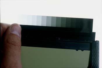
Figure 49 (above). A 4x5 dark slide modified by the inclusion of a transmission step wedge for reflection densitometric applications.
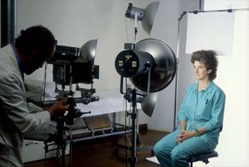
Figure 50 (above). A white light reflector above the subject as a control illuminant for imaging the step wedge onto film.
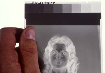
Figure 51 (above). The resulting negative with white illuminant and step wedge: allowing detailed analysis and compensation to be applied to densities measured. References
|
| © 2002 Prof. Robin Williams and Gigi Williams - Disclaimer URL: http://www.medicalphotography.com.au/Article_01/ Last modified: 3 May 2002 |