
about | articles | authors | contact | links
about | articles | authors | contact | links |
![]() Home > Articles > Fluorescence Photography > Applications of the Sodium Fluorescein technique
Home > Articles > Fluorescence Photography > Applications of the Sodium Fluorescein technique
FLUORESCENCE PHOTOGRAPHYAuthors: Prof. Robin Williams and Gigi Williams Applications of the Sodium Fluorescein technique:
|
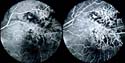 Figure 46. Fluorescein angiogram of the retina. |
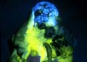 Figure 47. Turban Tumor and active blood supply. |
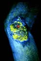 Figure 48. Carcinoma on knee and active blood supply. |
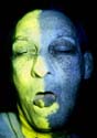 Figure 49. Blood supply to arteries. |
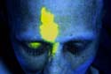 Figure 50. Distribution zone of supra-orbital artery. |
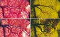 Figure 51. Fluorescein angiography of the brain. |
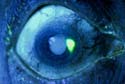 Figure 52. Delineation of a corneal ulcer. |
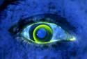 Figure 53. Poorly fitting contact lens. |
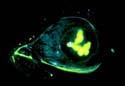 Figure 54. Delineation of corneal ulceration. |
| © 2002 Prof. Robin Williams and Gigi Williams - Disclaimer URL: http://www.medicalphotography.com.au/Article_02/ Last modified: 3 May 2002 |